Our Facilities

Computational Infrastructure

Computational biology is an interdisciplinary field that develops and applies computational techniques to solve biological problems. It entails close interaction between biological scientists, bioinformaticians, computer scientists, mathematicians, statisticians, physicists and researchers in other related disciplines. The role of computational biology has become increasingly central to developments in the biological sciences.

Within the ISMB, there are many computational biology PIs with access to dedicated computational resources, such as clusters and GPU servers. Key research areas include structural bioinformatics, genomics, molecular simulations and immunoinformatics.

Biophysics Centre

The ISMB Biophysics Centre provides access to instrumentation and expertise in the characterisation of biological macromolecules and their interactions, under the academic lead of Dr Mark Williams and with Centre Manager Nikos Pinotsis (n.pinotsis@bbk.ac.uk).

The ISMB Biophysics Centre provides access to instrumentation and expertise in the characterisation of biological macromolecules and their interactions, under the academic lead of Dr Mark Williams and with Centre Manager Dr Nikos Pinotsis.

A variety of instrumentation is available for the characterisation of equilibrium and kinetic behaviours of protein, nucleic acid and small-molecule interactions on a hire, service or collaborative basis, including:
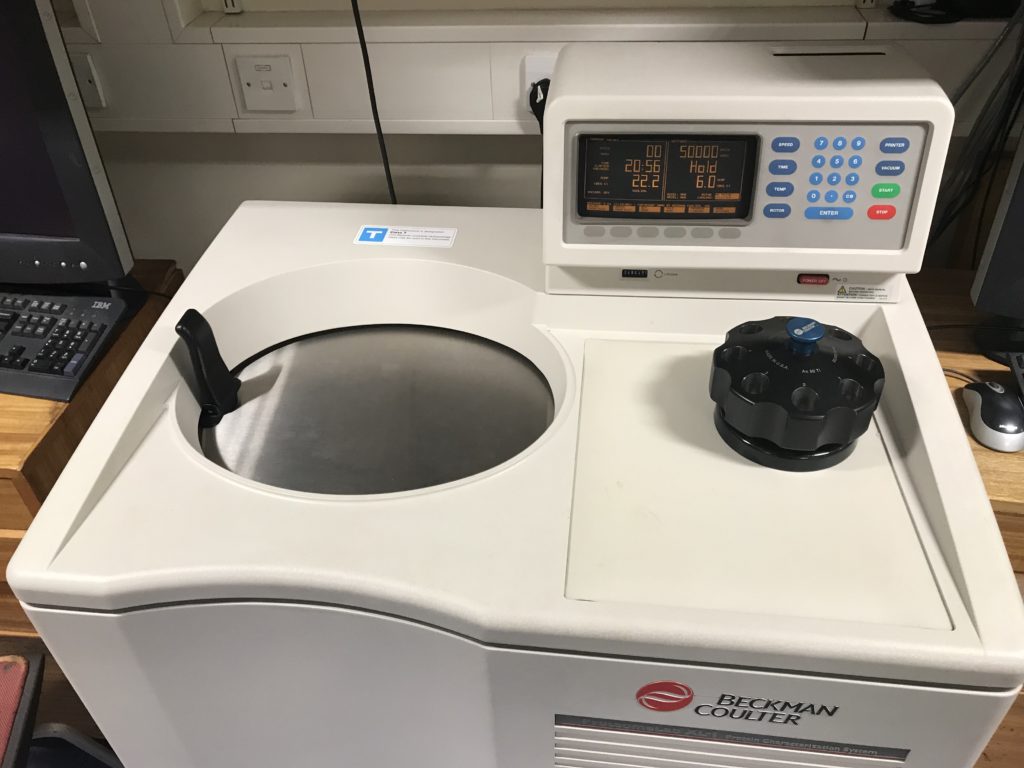
Researchers interested in using these technologies should contact the Biophysics Centre.

Trained users can book instruments though our online booking system.

Electron Microscopy

The renowned ISMB Electron Microscopy Lab was established by Professor Helen Saibil and is now headed by Professor Carolyn Moores, with Laboratory Co-ordinator Dr Natasha Lukoyanova and Laboratory Manager Dr Shu Chen. Researchers interested in using this technology should contact Dr Natasha Lukoyanova (n.lukoyanova@bbk.ac.uk)

Cryo-EM enables imaging of frozen-hydrated specimens in their native state without the need for dyes or fixatives, allowing the study of fine cellular structures, viruses and macromolecular complexes. Hardware and software advances have supported spectacular progress, so that the method is now often sufficient to determine structures to atomic resolution, and to sort out conformational variations.

The ISMB Electron Microscopy Lab was established by Professor Helen Saibil and is now headed by Professor Carolyn Moores, with Laboratory Co-ordinator Dr Natasha Lukoyanova and Laboratory Manager Dr Shu Chen. For single particle EM and tomography, the lab is equipped with Tecnai 100 kV, 120 kV, 200 kV FEG and 300 kV Krios G3i machines, with a K3 direct electron detector and BioQuantum energy filter, Falcon 3 detector and phase plate on the Krios, a DE20 direct detector on the F20, and a Vitrobot and Leica GP2 cryo plunger for sample preparation. Cell preparation equipment includes a high pressure freezer, cryo microtome and freeze substitution, as well as correlative cryo fluorescence microscopy/cryo EM.

Researchers interested in using this technology should contact Dr Natasha Lukoyanova.

More information is available on the EM lab page.

Birkbeck virtual EMBO course on cryo EM 2021. Apply now!

Birkbeck EMBO course on cryo EM 2019

Mass Spectrometry

Mass spectrometry is a powerful technique that become a key technology in structural molecular biology. The coupling of ion mobility to mass spectrometry has given rise to an even more powerful technique that can be used to report on the structure and dynamics of proteins and complexes of theoretically unlimited size. Researchers interested in using this technology should contact Dr Kostas Thalassinos (k.thalassinos@ucl.ac.uk).

Mass spectrometry is a powerful technique that is increasingly used in the field of structural molecular biology as it offers the following advantages:

  1.  High sensitivity, allowing the analysis of endogenously expressed proteins and protein complexes.
  2.  Ability to analyse several co-existing species present in a mixture.
  3. Monitoring of binding events and subunit exchange processes

Recently, mass spectrometry has been coupled to ion mobility spectrometry. Ion mobility is a gas-phase technique that separates ions based on their rotationally averaged collision cross-section (CCS) and charge. Ion mobility mass spectrometry offers additional advantages to mass spectrometry alone such as:

  1. The ability to distinguish between co-existing forms of a protein.
  2. Ability to study proteins of high flexibility.
  3. Experimental CCSs are in excellent agreement with those calculated from X-ray and NMR structures allowing the use of experimental CCSs to filter computer-generated models of oligomers.
  4. The ability to monitor and separate distinct conformational families co-existing in solution, such as in the case of oligomer formation

The ISMB houses a first generation Waters Synapt, the first commercially introduced ion mobility mass spectrometer. The Synapt in the ISMB has been modified for the analysis of very large ions such that analysis of protein complexes of up to 1 MDa are now possible.

Researchers interested in using this technology should contact Dr Kostas Thalassinos.

Molecular Interactions Facility

The UCL Molecular Interactions Facility provides access to protein purification equipment, advanced biophysical instrumentation, and computational modelling methods for the characterisation and structural determinations of biological macromolecules and their interactions, under the academic lead of Prof Steve Perkins (s.perkins@ucl.ac.uk) and Facility Manager Mr Jayesh Gor (j.gor@ucl.ac.uk).


The UCL Molecular Interactions Facility provides user access to protein purification, advanced biophysical instrumentation, and computational modelling for the characterisation and structural determinations of biological macromolecules and their interactions. Together these techniques are advantageous in generating molecular structures in solution that complement those determined by crystallography, NMR or cryo-EM. The costs of annual service contracts are met from access charges from users. We lead the CCP-SAS project for atomistic scattering modelling in the UK (http://www.ccpsas.org/) that started in 2013 and has over 50 publications to its credit.

  • AKTA protein purification apparatus
  • Analytical ultracentrifugation (two instruments, one equipped with fluorescent optics)
  • Dual polarisation interferometry
  • Neutron scattering (at the ISIS and ILL facilities)
  • Molecular dynamics and Monte Carlo simulations of solution scattering data
  • Surface plasmon resonance (Biacore X-100)
  • X-ray scattering (at the Diamond Light Source and the ESRF)

Researchers interested in these approaches should contact Professor Steve Perkins and the Facility Manager Jayesh Gor.

In addition, the ISMB Biophysics Centre provides access to a large range of instrumentation for macromolecular characterisation and functional studies.


Nuclear Magnetic Resonance

The ISMB NMR facility aims to expand the frontiers of biomolecular NMR spectroscopy through a combination of advanced method development and their application to important questions in modern biology. Particular expertise exists in understanding protein dynamics: NMR can report with exquisite sensitivity and at residue specific level on biological motions across a range of timescales from picoseconds to days. Researchers interested in using this technology should contact Dr Angelo Figueiredo (m.figu@ucl.ac.uk).

The ISMB NMR facility aims to expand the frontiers of biomolecular NMR spectroscopy through a combination of advanced method development and their application to important questions in modern biology. Particular expertise exists in understanding protein dynamics: NMR can report with exquisite sensitivity and at residue specific level on biological motions across a range of timescales from picoseconds to days.

Thanks to recent support from the Wellcome Trust through a Multi-User Equipment Grant led by NMR Facility Director Professor John Christodoulou and with Professor Flemming Hansen, the ISMB now holds three NMR spectrometers ranging in magnetic field from 11.7 Tesla to 18.8 Tesla with high-sensitivity cryogenic radio-frequency probes installed for each. An experienced NMR spectroscopist (Dr Angelo Figuerido) has overseen the recent upgrade and manages the facility.

Researchers interested in using this technology should contact Dr Angelo Figueiredo.

Single Molecule Biophysics

Single-molecule techniques enable the manipulation and visualisation of biological processes at the single-molecule level. They are at the frontier of structural biology as they enable high-resolution measurements of the dynamics, kinetics and mechanics that underpin biomolecular interactions. These techniques enable researchers to achieve the following:

(i) Measure changes in distance (with sub-nanometer resolution), providing detailed insight into structural transitions

(ii) Probe biomolecular interactions in real-time (with millisecond resolution), allowing the measurement of highly dynamic processes

(iii) Apply and/or measure mechanical force (on the scale of picoNewtons), allowing researchers to (a) probe the impact of forces generated in the cell and (b) study the mechanical stability of biomolecules

(iv) Identify rare events and states that would otherwise be obscured in standard bulk measurements

(v) Visualise structures below the diffraction limit (~250 nm) using high-resolution single-molecule imaging


The laboratory of Dr Graeme King houses a state-of-the art dual-trap optical tweezers instrument (LUMICKS M-trap). This allows real-time manipulation of single molecules (such as DNA and proteins) as well as measure the length of the molecule and the forces acting on it with sub-nanometer and sub-picoNewton resolution. The M-trap is is integrated with a multi-channel microfluidics system, which enables researchers to control the stepwise assembly of molecular complexes and rapidly change the environment of the molecule. The King lab has expertise in the use of this technology to study the mechanisms and functions of DNA and protein interactions.

The laboratory of Dr Alan Lowe has constructed instruments with the following capabilities:

1) Single-molecule Forster Resonance Energy Transfer (smFRET)
2) Single-molecule Tracking
3) Super-resolution Localisation Microscopy (dSTORM and PALM)
4) Timelapse imaging
5) Total Internal Reflection Fluorescence (TIRF) imaging

Researchers interested in using the above instruments should contact Dr Graeme King [link to webpage] or Dr Alan Lowe [link to webpage].

X-Ray Crystallography

The ISMB X-Ray Crystallography Laboratory was completely refurbished in 2011 and houses state-of-the-art equipment for successful screening and optimization of crystallographic experiments. Researchers interested in using this technology should contact the ISMB X-Ray Crystallography Lab Manager Nikos Pinotsis (n.pinotsis@bbk.ac.uk).


The ISMB X-Ray Crystallography Laboratory was completely refurbished in 2011 and houses state-of-the-art equipment for successful screening and optimization of crystallographic experiments. This research facility aims to provide a self-contained service to members of the ISMB, including:

  • Robotics facilities for large-scale screening and production of de novo optimization screens.
  • Facilities for manual setup, as well as crystal manipulation and freezing.
  • Two temperature-controlled rooms: one at 4°C and the other at 20°C (the latter with automatic crystal storage and imaging systems for crystal detection).
  • A facility for in-house data collection using a modern Rigaku detector with CCD image collection equipment.